Antibody Identity card
Antibody information [IL4I1 ]
1006231
- Clone name 1006231
- Description Rat monoclonal
- Antigen used CHO derived hIL4i1 Met1-His567
- Epitope Information not available
- Isotype Rat IgG2a
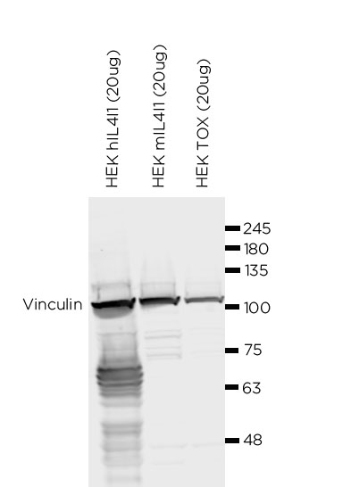
- Confirmed species reactivity Human
- Ab used R&D Systems (cat MAB56843) 0.5 mg/ml
| APPLICATION | Recommended concentration | Status | Protocol |
|---|---|---|---|
| Western blotting (WB) | 2 ug/mL | Working | Western Blotting (WB) |
| Immunocytochemistry (ICC) | Neat tissue culture supernatant | Working | ICC in frozen tissue and cytospin preparation |
| Immunohistochemistry (IHC-P) | 0.41 ug/ml | Working | |
| Immunoflourescence (IF) | 1.25 ug/ml | Working | Immunofluorescence staining |
| Flow cytometry (FC) | 2.5 ug/ml | Working | Flow cytometry |
| IHC-P Species | Not tested |
Ab ID Card application validation / characterisation
WB Validation
Over-expression/cross reactivity
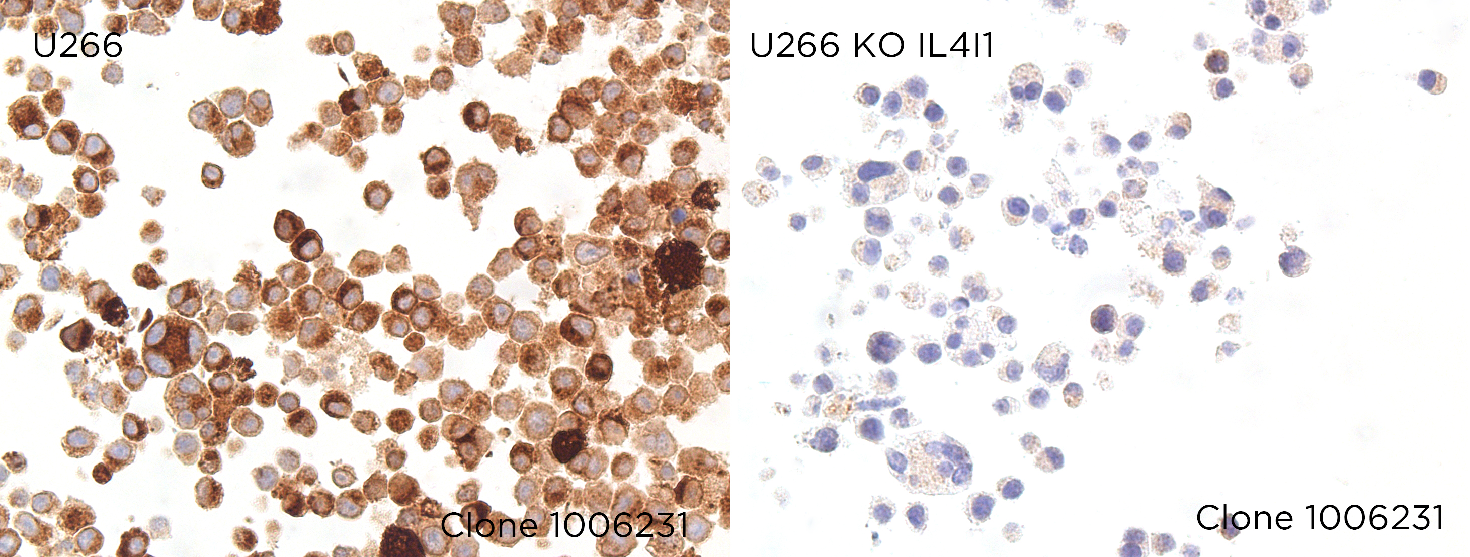
Gene inactivation
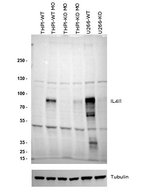
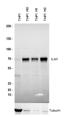
WB Characterisation
Endogenous expression
ICC / ICC-P Validation
Over-expression/cross reactivity
Gene inactivation
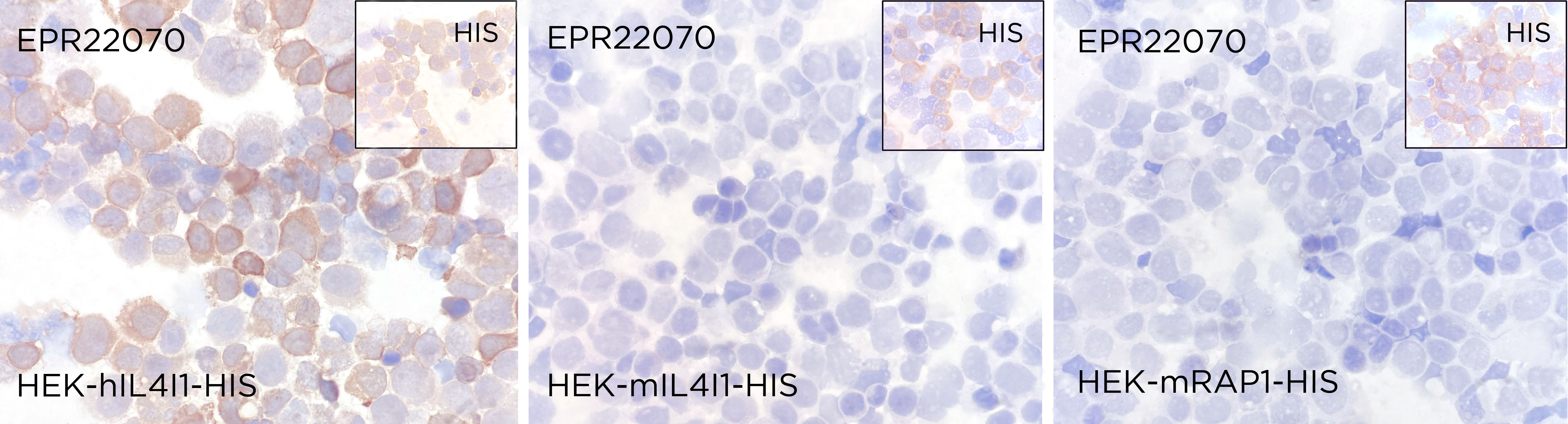
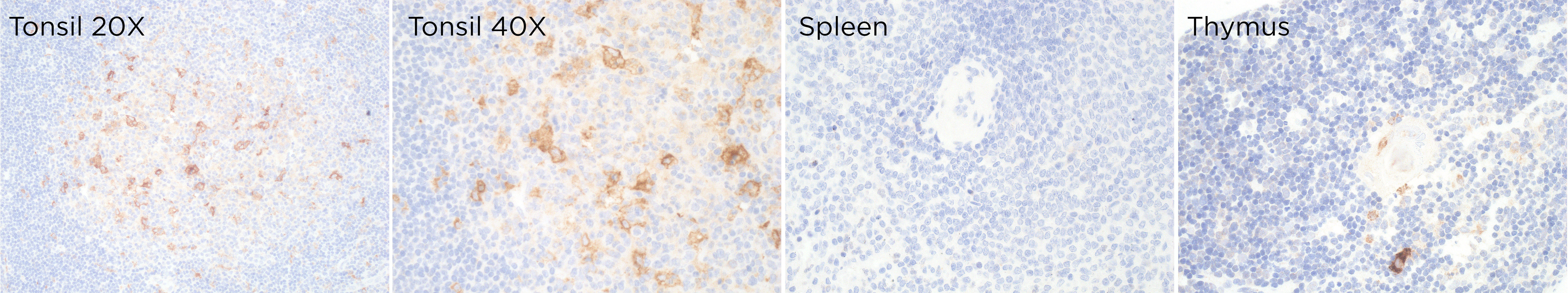
IHC-P Characterisation
Endogenous expression
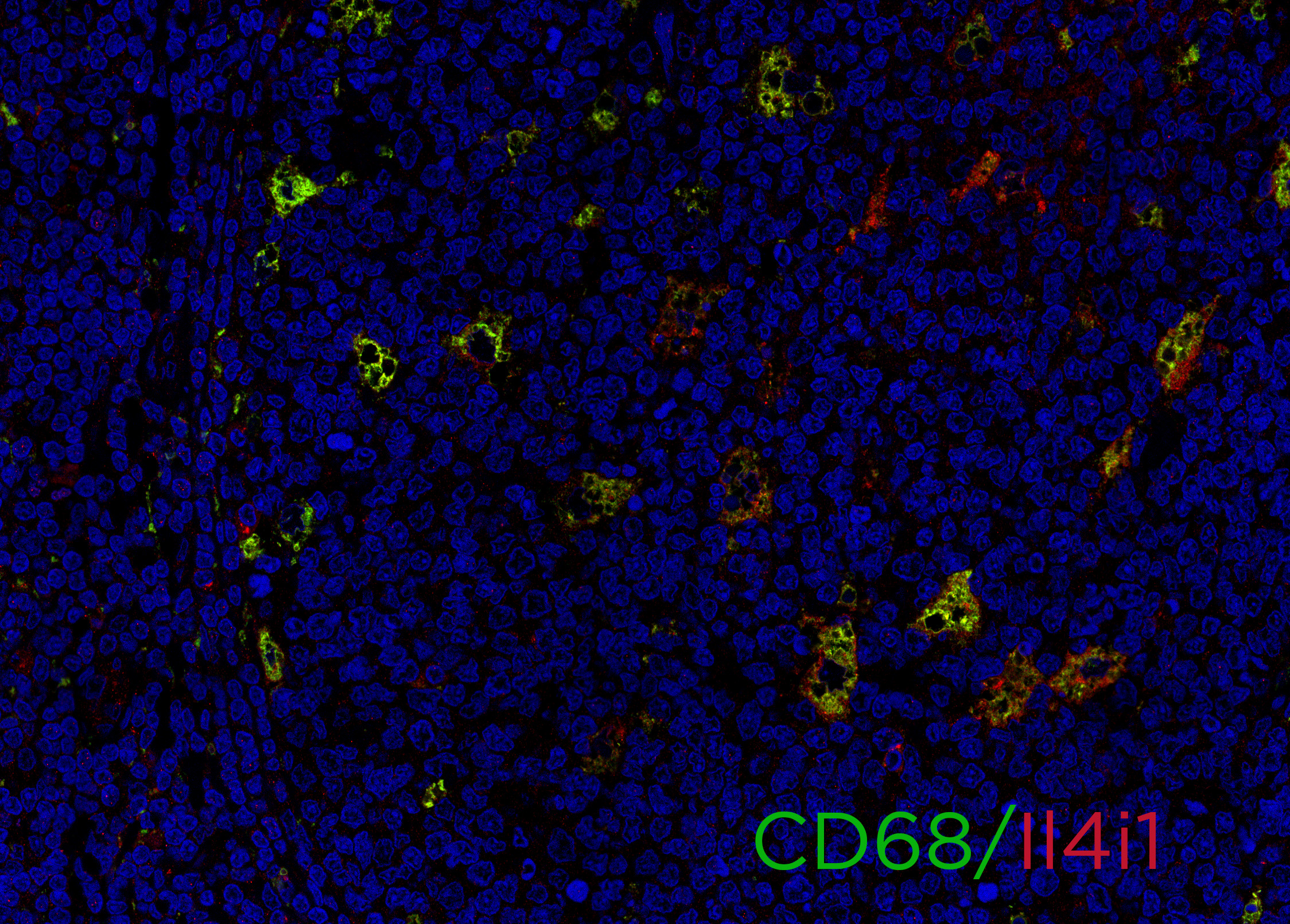
IF Characterisation
Endogenous expression
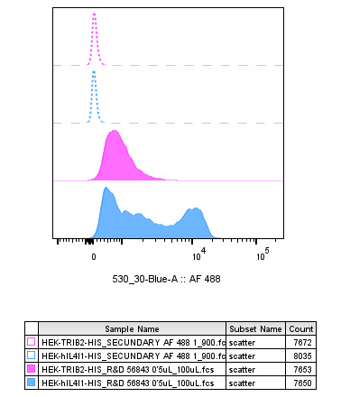
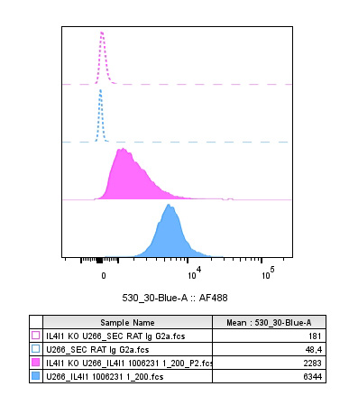
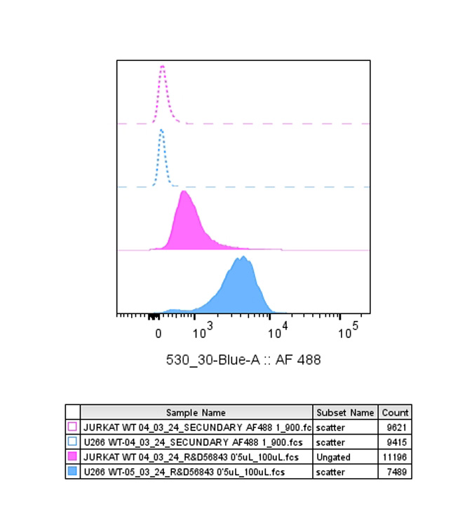
FC Validation
Over-expression/cross reactivity
Gene inactivation
FC Characterisation
Endogenous expression
IHC-P Domestic species
Not tested
IHC-P Wild species
Not tested