Antibody Identity card
Antibody information [IL4I1 ]
BALI82E
- Clone name BALI82E
- Description Rat monoclonal
- Antigen used Human IL4I1-His-Strep expressed in 293 cells
- Epitope Unknown
- Isotype IgG2a, K
- Confirmed species reactivity Human
- Ab used Tissue culture supernatant
| APPLICATION | Recommended concentration | Status | Protocol |
|---|---|---|---|
| Western blotting (WB) | Tissue culture supernatant | Working | Western Blotting (WB) |
| Immunocytochemistry (ICC) | Neat tissue culture supernatant | Working | ICC in frozen tissue and cytospin preparation |
| Immunohistochemistry (IHC-P) | 1:10 Neat tissue culture supernatant | Working | BOND-MAX automated immunohistochemistry |
| Immunoflourescence (IF) | 1:2 Neat tissue culture supernatant | Working | Immunofluorescence staining |
| Flow cytometry (FC) | 40ul culture supernatant/tube | Not working | Flow cytometry |
| IHC-P Species | Not tested |
Ab ID Card application validation / characterisation
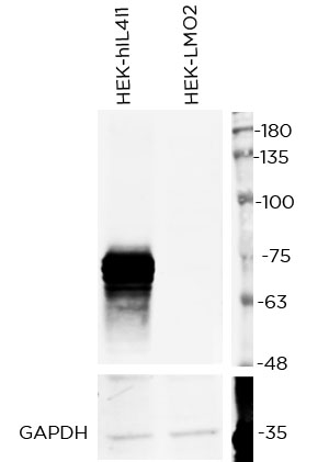
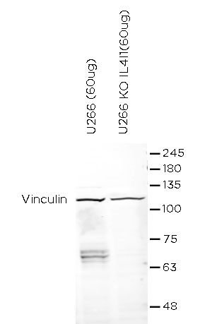
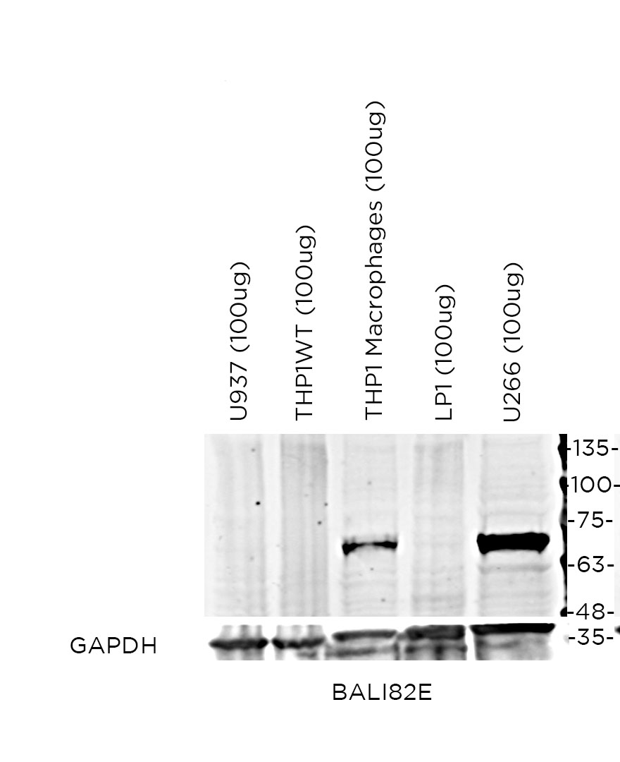
WB Validation
Over-expression/cross reactivity
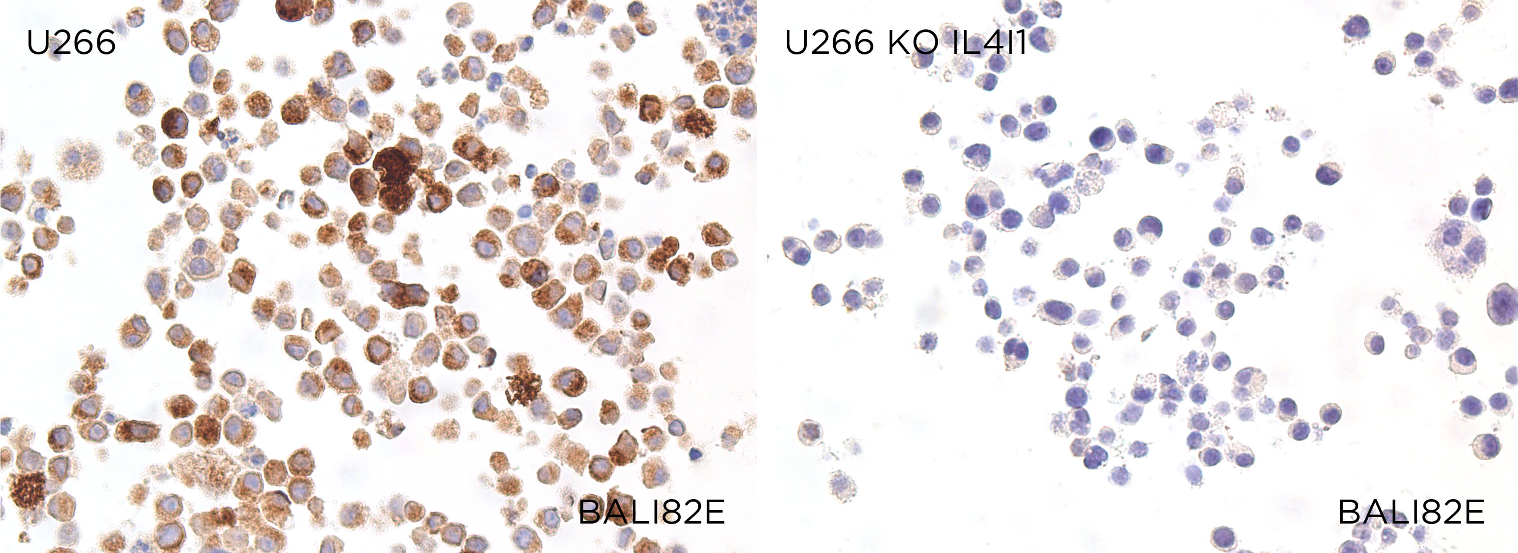
Gene inactivation
WB Characterisation
Endogenous expression
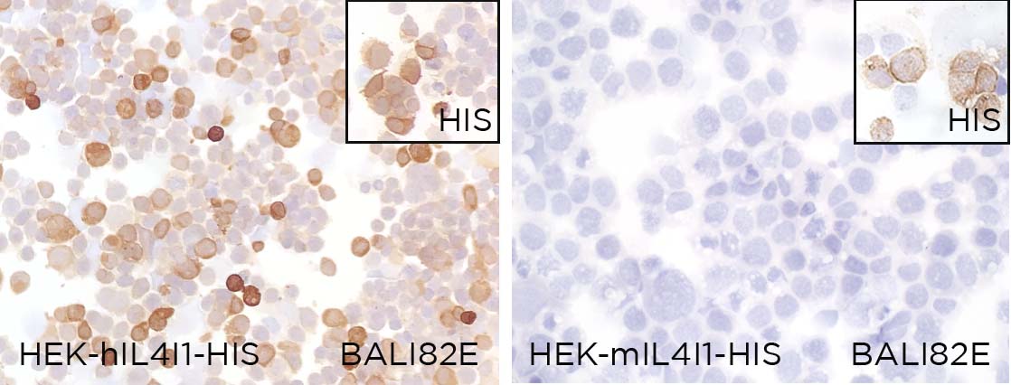
ICC / ICC-P Validation
Over-expression/cross reactivity
Gene inactivation
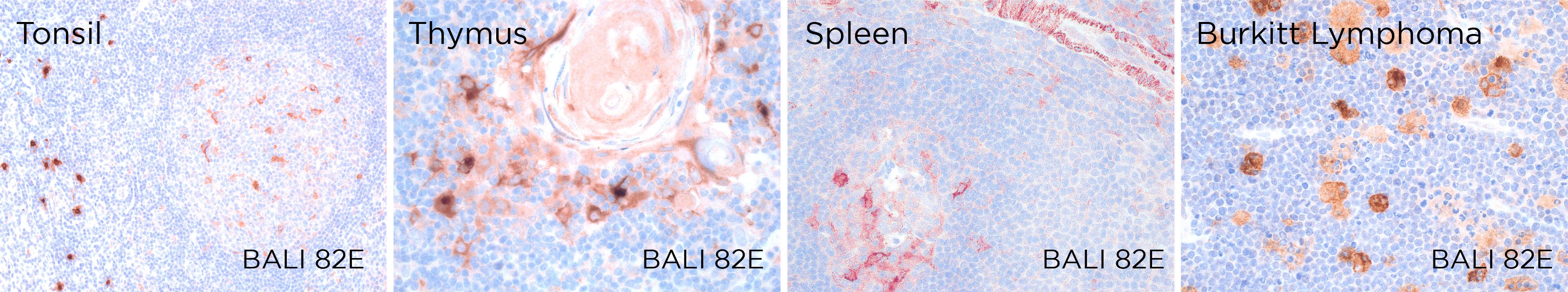
IHC-P Characterisation
Endogenous expression
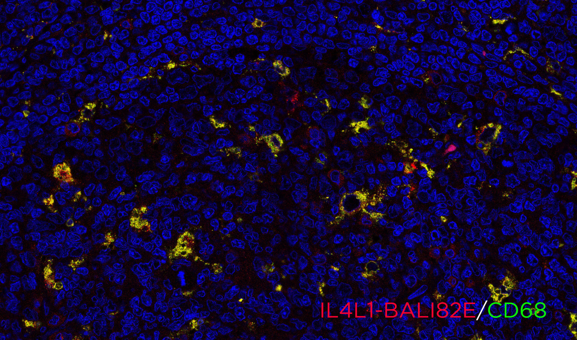
IF Characterisation
Endogenous expression
FC Validation
Over-expression/cross reactivity
Not working
Gene inactivation
Not working
FC Characterisation
Endogenous expression
Not working
IHC-P Domestic species
Not tested
IHC-P Wild species
Not tested