Antibody Identity card
Antibody information [IL4I1 ]
EPR22070
- Clone name EPR22070
- Description Rabbit monoclonal
- Antigen used Information not available
- Epitope
- Isotype IgG
- Confirmed species reactivity Human
- Ab used Anti-IL-4I1/LAO Abcam (Cat. ab222102) 0.567mg/ml
| APPLICATION | Recommended concentration | Status | Protocol |
|---|---|---|---|
| Western blotting (WB) | 0.567 μg/ml | Working | Western Blotting (WB) |
| Immunocytochemistry (ICC) | 0.567 μg/ml | Working | ICC in frozen tissue and cytospin preparation |
| Immunohistochemistry (IHC-P) | 0.567 μg/ml | Working | BOND-MAX automated immunohistochemistry |
| Immunoflourescence (IF) | 1.134 μg/ml | Working | Immunofluorescence staining |
| Flow cytometry (FC) | 0.945 ug/ml | Working | Flow cytometry |
| IHC-P Species | Not tested |
Ab ID Card application validation / characterisation
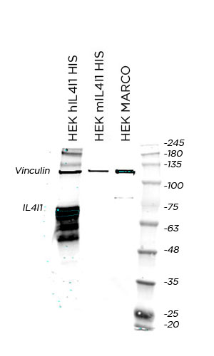
WB Validation
Over-expression/cross reactivity
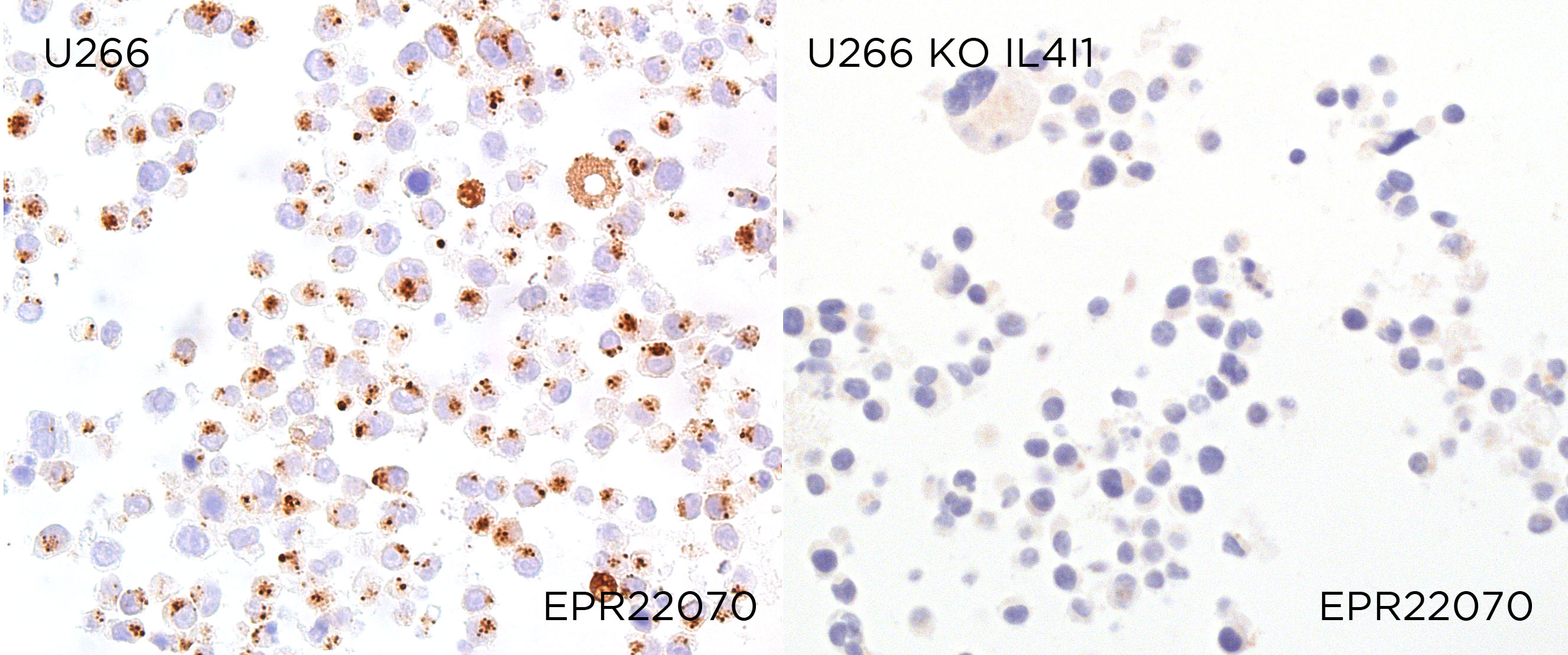
Gene inactivation
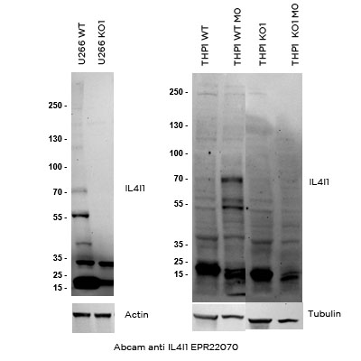
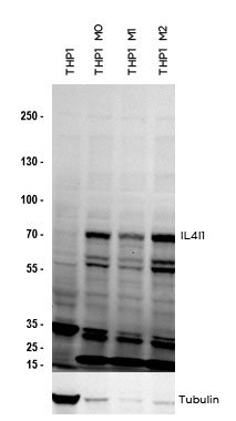
WB Characterisation
Endogenous expression
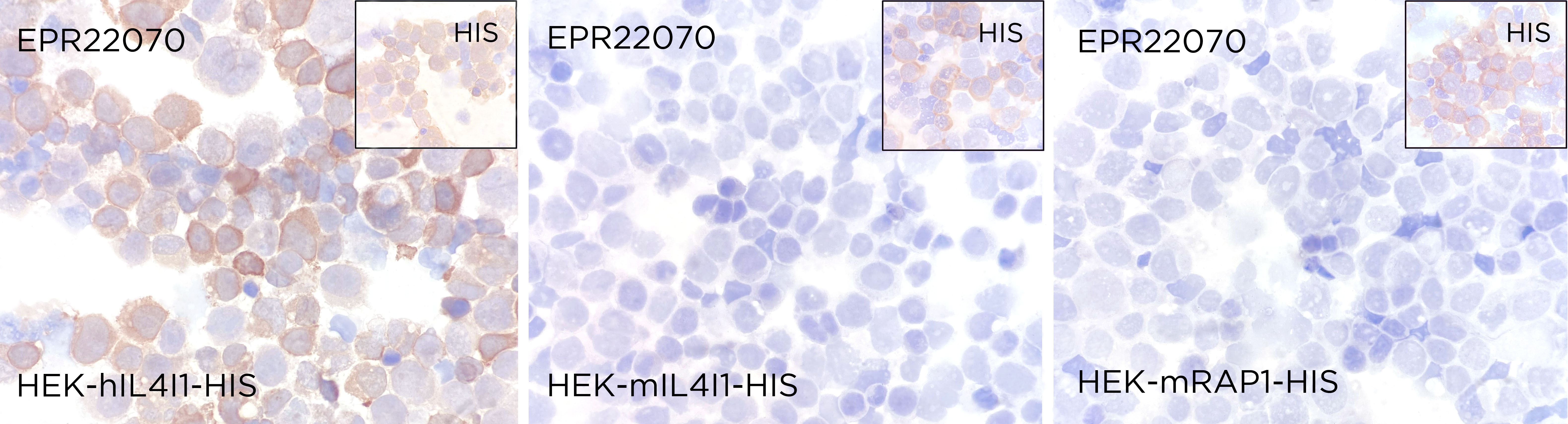
ICC / ICC-P Validation
Over-expression/cross reactivity
Gene inactivation
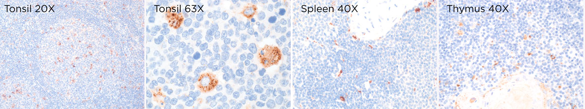
IHC-P Characterisation
Endogenous expression
IF Characterisation
Endogenous expression

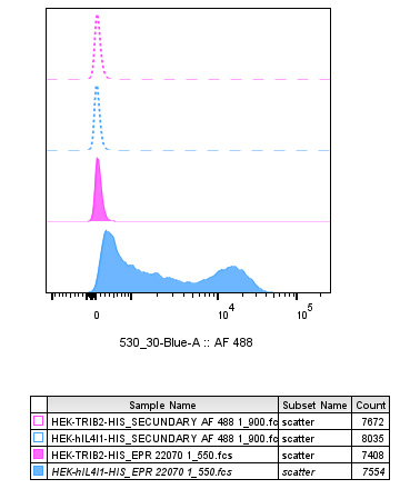
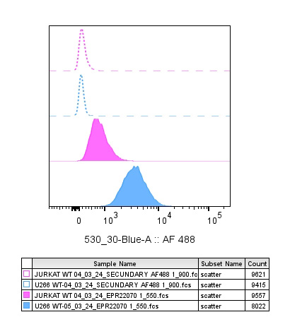
FC Validation
Over-expression/cross reactivity
Gene inactivation
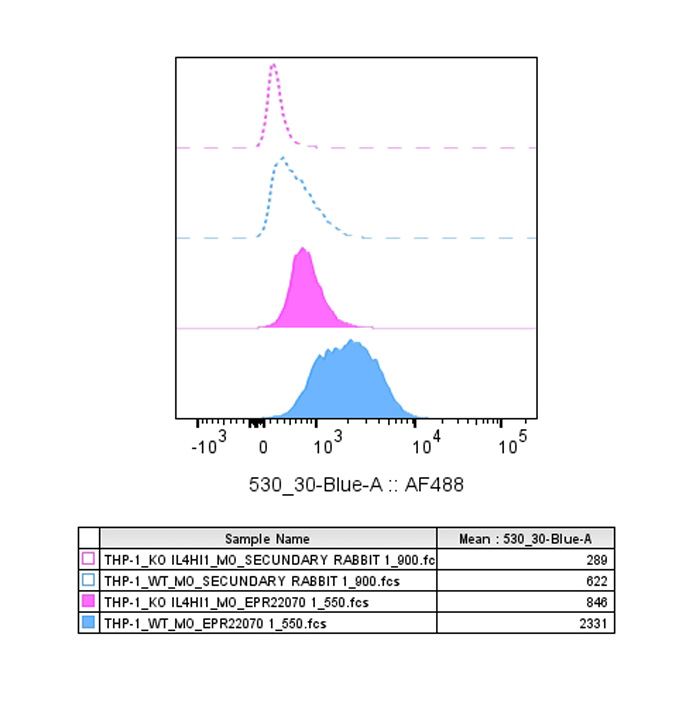
FC Characterisation
Endogenous expression
IHC-P Domestic species
Not tested
IHC-P Wild species
Not tested