Antibody Identity card
Antibody information [MNDA]
5C
- Clone name 5C
- Description Mouse monoclonal
- Antigen used Full length MNDA-GST recombinant protein
- Epitope Unknown
- Isotype IgG1
- Confirmed species reactivity Human
- Ab used Purified antibody (5C) CNIO (2.27 mg/ml)
| APPLICATION | Recommended concentration | Status | Protocol |
|---|---|---|---|
| Western blotting (WB) | 22.7 ug/ml | Working | Western Blotting (WB) |
| Immunocytochemistry (ICC) | 5.7 ug/ml | Working | ICC in frozen tissue and cytospin preparation |
| Immunohistochemistry (IHC-P) | 22.7 ug/ml | Working | BOND-MAX automated immunohistochemistry |
| Immunoflourescence (IF) | 37.83 ug/ml | Working | Immunofluorescence staining |
| Flow cytometry (FC) | 2.27 ug/ml | Working | Flow cytometry |
| IHC-P Species | Not tested |
Ab ID Card application validation / characterisation
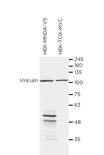
WB Validation
Over-expression/cross reactivity
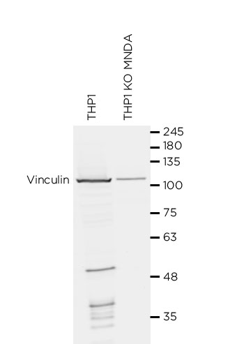
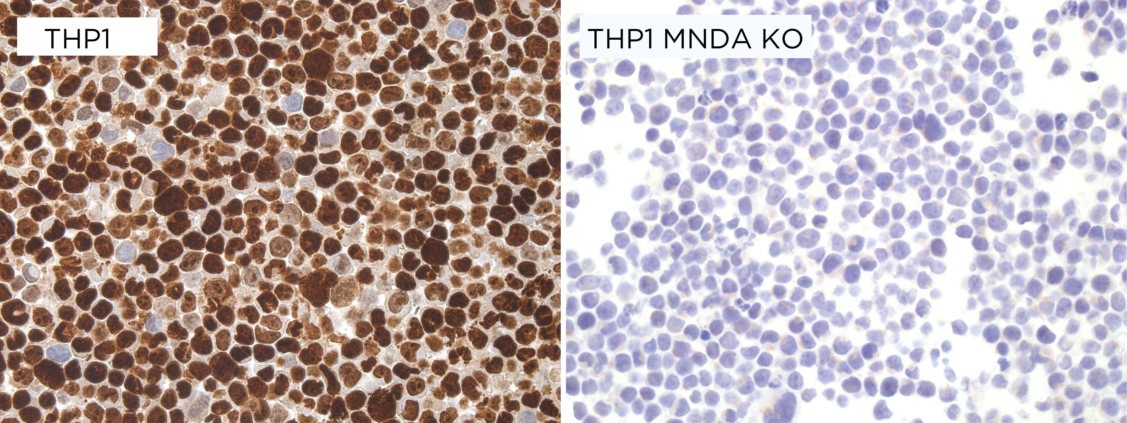
Gene inactivation
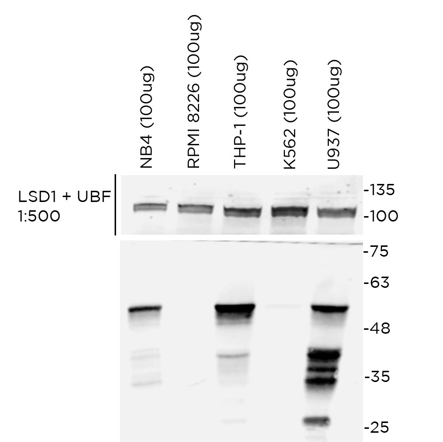
WB Characterisation
Endogenous expression
ICC / ICC-P Validation
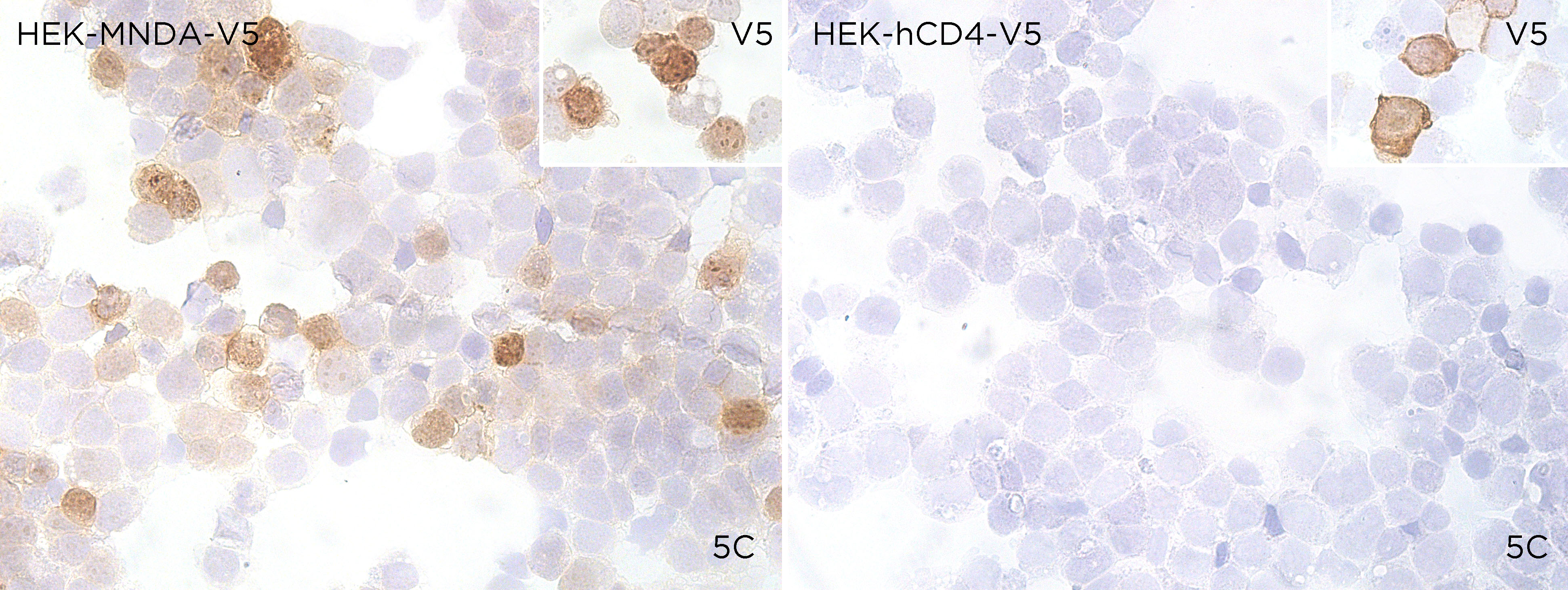
Over-expression/cross reactivity
Gene inactivation
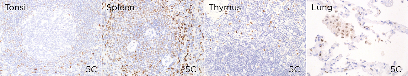
IHC-P Characterisation
Endogenous expression
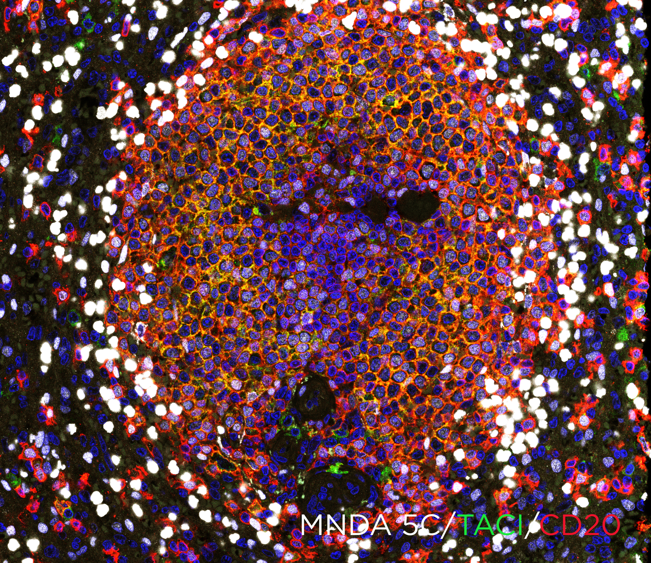
IF Characterisation
Endogenous expression
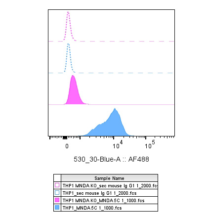
FC Validation
Over-expression/cross reactivity
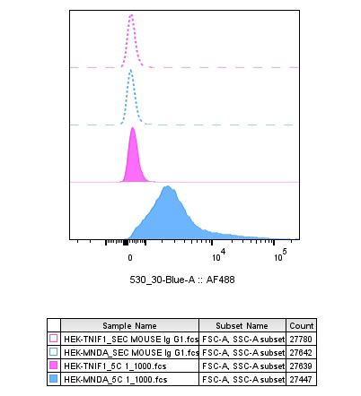
Gene inactivation
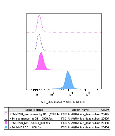
FC Characterisation
Endogenous expression
IHC-P Domestic species
Not tested
IHC-P Wild species
Not tested