mAbs Against HUMAN Protein
HP-1α
| STATUS: |  |
|---|---|
| CONTACT INFORMATION: | Monoclonal Antibodies Unit, Centro Nacional de Investigaciones Oncológicas |
| STATUS: | Validated |
| TYPE: | rat anti human |
| CLONE NAME: | HP330A |
| PROTEIN: | human full length HP1α |
| PROTEIN WEB: | http://www.hprd.org/summary?hprd_id=05131&isoform_id=05131_3&isoform_name=Isoform_1 |
| ANTIGEN USED: | GST-HP1α and GST-HP1γ recombinant proteins |
| FUSION PARTNER: | NS-1 |
| ISOTYPE: | IgG2a |
| SPECIES REACTIVITY: | Human |
| PREPARATION AND STORAGE: | Aliquot and store at 4C. Do not freeze |
| APP RECOMMENDED: | IHQ-paraffin, IHQ-frozen, IF, WB |
| APP NO TESTED: | IP, Flow cytometry |
Description
Heterochromatin protein-1 (HP1) is a methyl-lysine binding protein localized at heterochromatin sites, where it mediates gene
silencing. HP-1alpha is involved inthe formation of functional kinetochore through interactionwith MIS12 complex proteins. HP-
1alpha also interacts with lamin B receptor, contributing to the association of the heterochromatin with the inner nuclear membrane.
Applications
| IHC Techniques | Clone | Dilution | Antibody concentration | Antigen retrieval method | Visualization kit | +/- control | Protein localization | Positivity in other species | Protocol |
|---|---|---|---|---|---|---|---|---|---|
| Frozen tissue and cytospins | |||||||||
Recommended Result obtained is satisfactory. The reagent can be use in this application | HP330A | 1:20 | supernatant | None | rabbit anti rat HRP | Tonsil / | Nuclear | not done | |
| Paraffin tissue | |||||||||
Recommended Result obtained is satisfactory. The reagent can be use in this application | HP330A | 1:20 | upernatant | 20 min ER2 | Tonsil / | Nuclear | not done | Available | |
| Immunofluorescence | |||||||||
Recommended Result obtained is satisfactory. The reagent can be use in this application | HP330A | neat | upernatant | 20 min ER2 | Tonsil / | Nuclear | not done | Available | |
 Enlarge image
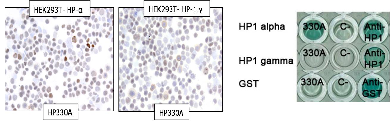
- HP1 (330A) IHC in transfected cytospins and by ELISA
- Heterochromatin protein-1 (HP1) is a methyl-lysine binding protein localized at heterochromatin sites and expressed in all cell types. We made a mAb against HP1 protein using, as immunogen, a combination of HP1 alpha and HP1 gamma protein. We could not distinguish by IHC in tranfected Hek HP1 alpha and HP1 gamma which subunit was recognized by our antibody since strong nuclear staining was observed in both cytospins. Using ELISA technique we could determine that our antibody is specific for the alpha subunit.
 Enlarge image
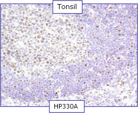
- Rat monoclonal 330A can be used to detect HP1 α expression in frozen human tissues.
 Enlarge image
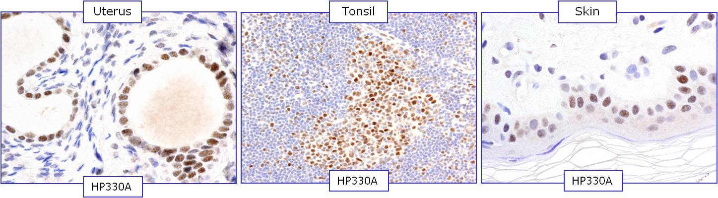
- Rat monoclonal 330A can be used to detect HP1 α expression in paraffin human tissue
 Enlarge image
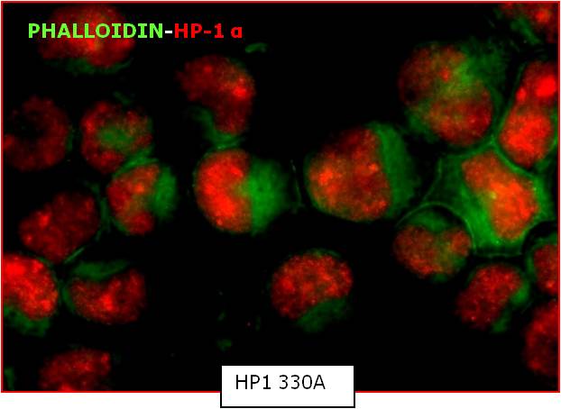
- Immunofluorescence of HP1 (330A) mAb in Ramos cell line
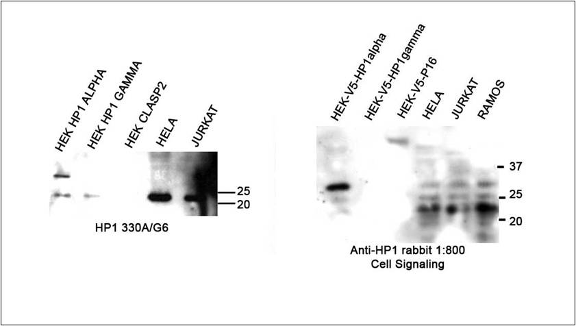
| WB Techniques | Clone | Dilution | Antibody concentration | +/- control | Expected MW | Observed Mw | Positivity in other species | Protocol |
|---|---|---|---|---|---|---|---|---|
| Western Blotting | ||||||||
Recommended Result obtained is satisfactory. The reagent can be use in this application | HP330A | neat | supernatant | Hela / | 22kDa | 22kDa | Not done | Available |
| Immunoprecipitation | ||||||||
 Enlarge image
- Rat monoclonal HP330A can be used to detect HP1α protein in human cell lines by WB.
- Lanes
Lane 1 Transfected Hek-V5-HP1 α (10цg) (+)
Lane 2 Transfected Hek-V5-HP-1 γ (10цg) (-)
Lane 3 Transfected Hek-V5-P16 (10цg) (-)
Lane 4 Hela cell line (100цg) (+)
Lane 5 Jurkat cell line (100цg) (+)
Lane 6 Ramos cell line (100 цg) (+)
Rabbit antibody from Cell Signaling was used at 1:800