Antibody Identity card
Antibody information [ PDL-1 mouse specific]
EPR20529
- Clone name EPR20529
- Description Rabbit monoclonal
- Antigen used Recombinant fragment
- Epitope Unknown
- Isotype IgG
- Confirmed species reactivity Mouse
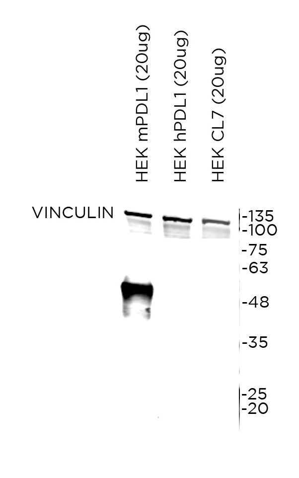
- Ab used Abcam Anti-PD-L1 (ab213480) 0.508mg/ml
| APPLICATION | Recommended concentration | Status | Protocol |
|---|---|---|---|
| Western blotting (WB) | 1 ug/ml | Working | Odyssey Western Blotting protocol (OdWB) |
| Immunocytochemistry (ICC) | 0.25 ug/ml | Working | ICC in frozen tissue and cytospin preparation |
| Immunohistochemistry (IHC-P) | 0.25 ug/ml | Working | PDL-1 mAb IHC-P in mouse tissues |
| Immunoflourescence (IF) | Not tested | Immunofluorescence staining | |
| Flow cytometry (FC) | 1.016 ug/ml | Working | Flow cytometry |
| IHC-P Species | Not tested |
Ab ID Card application validation / characterisation
WB Validation
Over-expression/cross reactivity
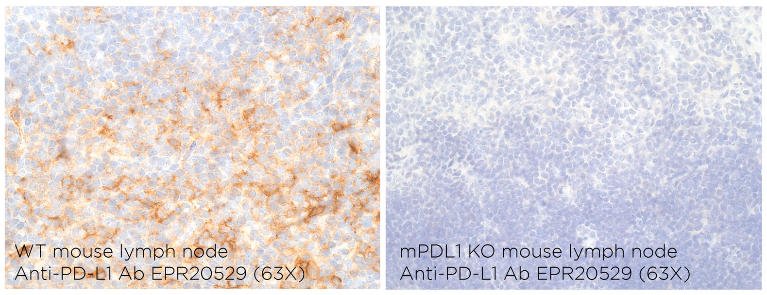
Gene inactivation
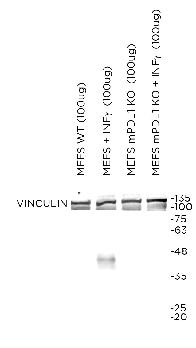
WB Characterisation
Endogenous expression
ICC / ICC-P Validation
Over-expression/cross reactivity
Gene inactivation
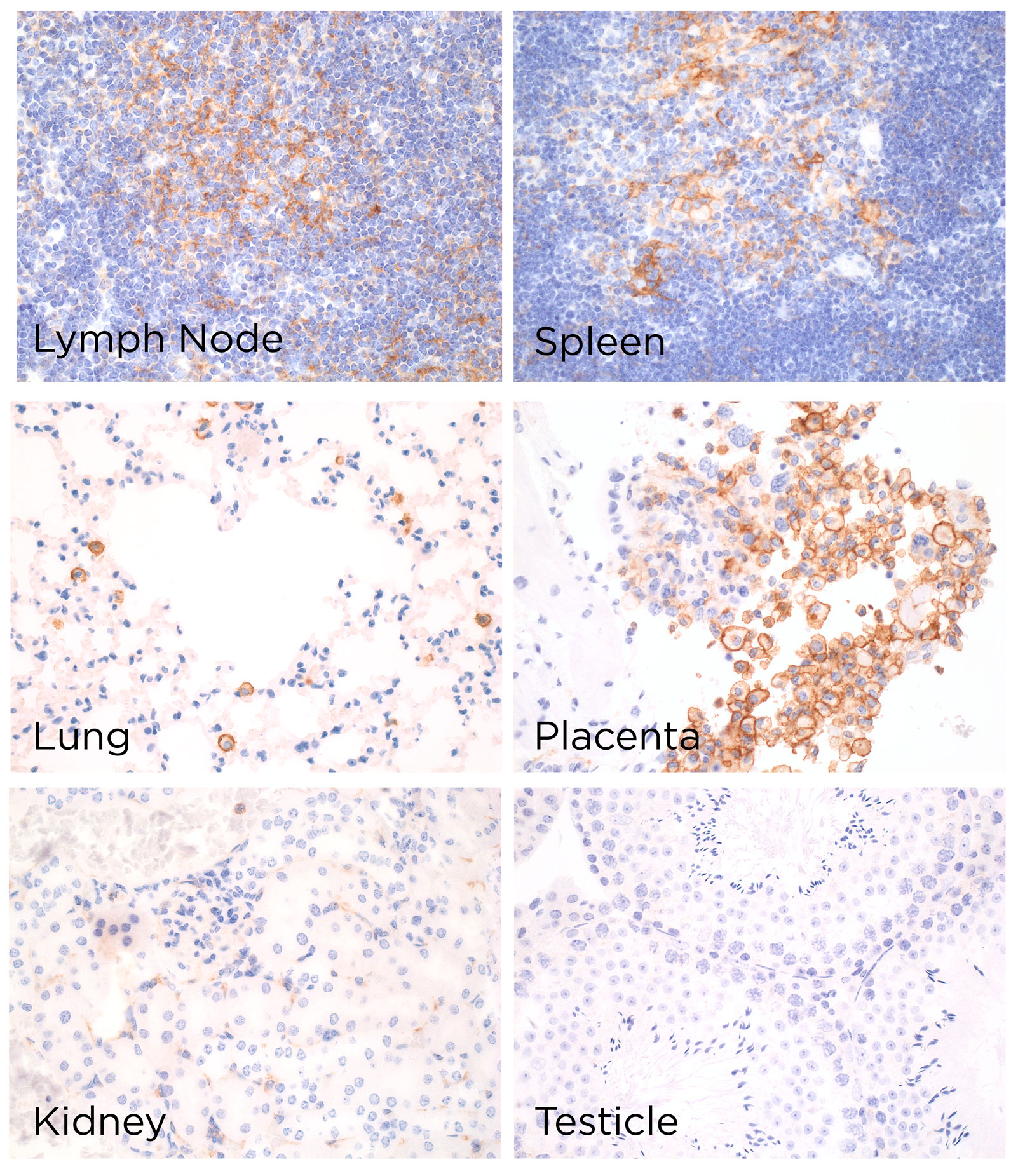
IHC-P Characterisation
Endogenous expression
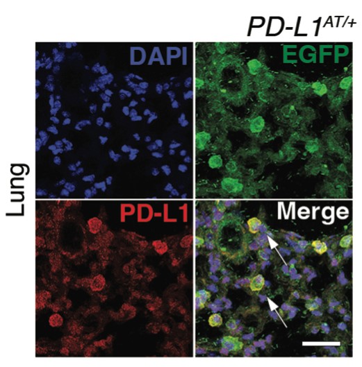
IF Characterisation
Endogenous expression
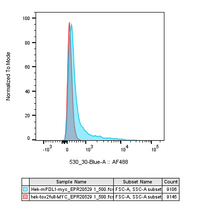
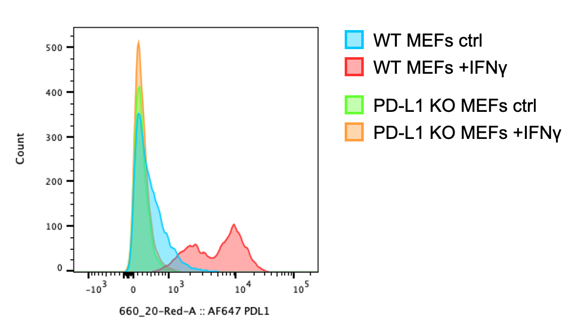
FC Validation
Over-expression/cross reactivity
Gene inactivation
FC Characterisation
Endogenous expression
Not tested
IHC-P Domestic species
Not tested
IHC-P Wild species
Not tested