Antibody Identity card
Antibody information [TOX2]
TOM924D
- Clone name TOM924D
- Description Rat monoclonal
- Antigen used HIS-SUMO-hTOX2 tv2 (full length)-Strep-tag2
- Epitope VVEAGK
- Isotype IgG2a
- Confirmed species reactivity Human, mouse
- Ab used CNIO purified antibody (concentration 3.94 mg/ml)
| APPLICATION | Recommended concentration | Status | Protocol |
|---|---|---|---|
| Western blotting (WB) | 7ug/ml | Working | Odyssey Western Blotting protocol (OdWB) |
| Immunocytochemistry (ICC) | 3.94 ug/ml | Working | ICC in frozen tissue and cytospin preparation |
| Immunohistochemistry (IHC-P) | 0.98 ug/ml | Working | BOND-MAX automated immunohistochemistry |
| Immunoflourescence (IF) | 1.97 ug/ml | Working | Immunofluorescence staining |
| Flow cytometry (FC) | 0.985 ug/ml | Not working | Flow cytometry |
| IHC-P Species | 3.94 μg/ml | Working |
Ab ID Card application validation / characterisation
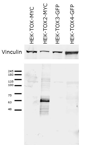
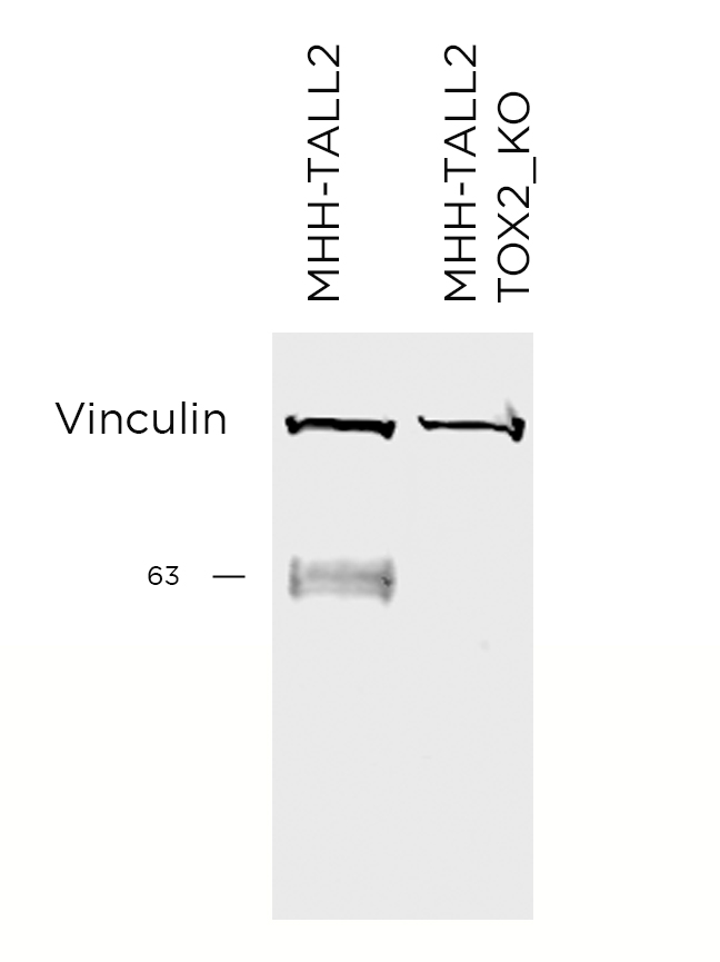
WB Validation
Over-expression/cross reactivity
Gene inactivation
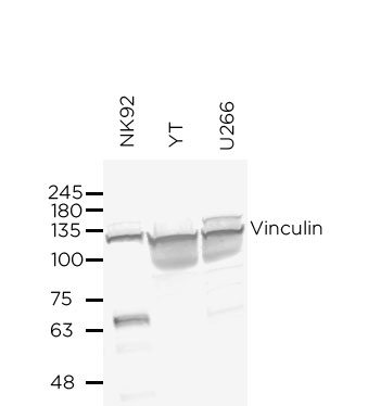
WB Characterisation
Endogenous expression
ICC / ICC-P Validation
Over-expression/cross reactivity
Gene inactivation
IHC-P Characterisation
Endogenous expression
IF Characterisation
Endogenous expression
FC Validation
Over-expression/cross reactivity
Not working
Gene inactivation
Not working
FC Characterisation
Endogenous expression
Not working
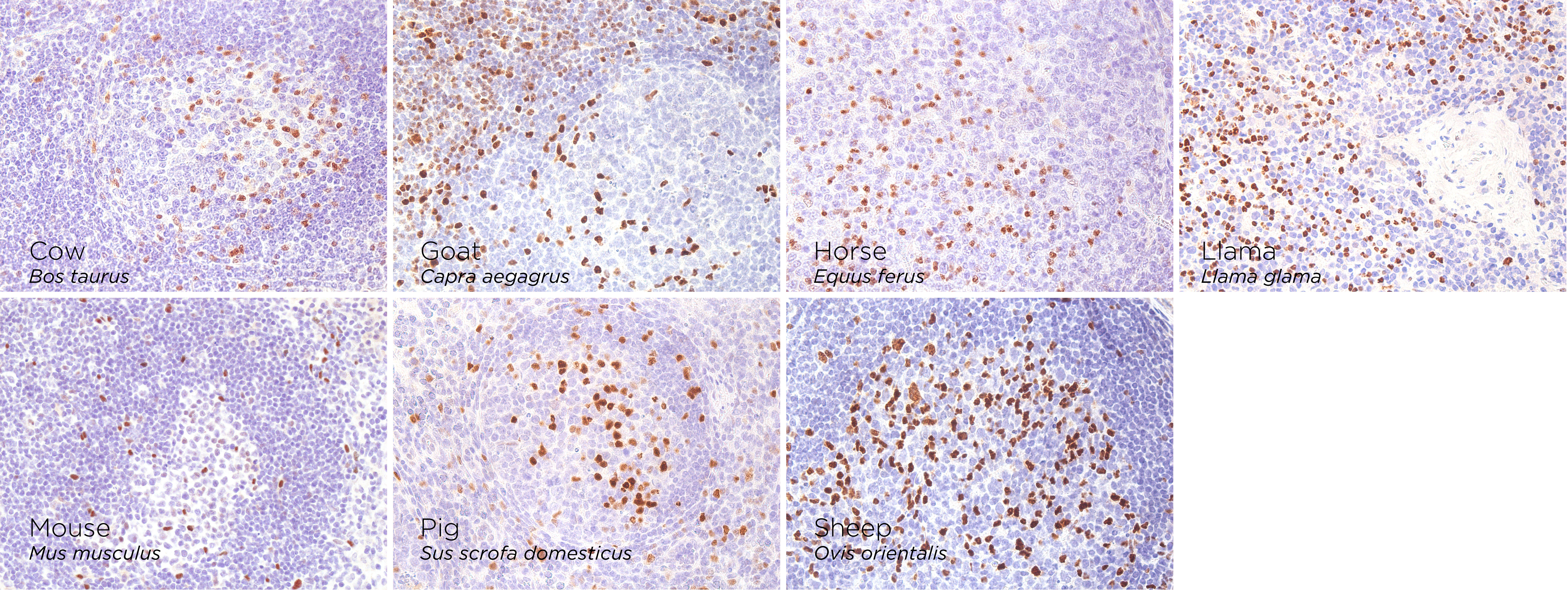
IHC-P Domestic species
- Canary Serinus canaria
- -
- Cat Felis catus
- -
- Cow Bos taurus
- +++
- Dog Canis lupus familiaris
- -
- Goat Capra aegagrus
- +++
- Horse Equus ferus
- ++
- Jacobin Pigeon Columba livia
- -
- Llama Llama glama
- +++
- Mouse Mus musculus
- +++
- Pig Sus scrofa domesticus
- +++
- Rat Rattus norvegicus
- -
- Sheep Ovis orientalis
- +++
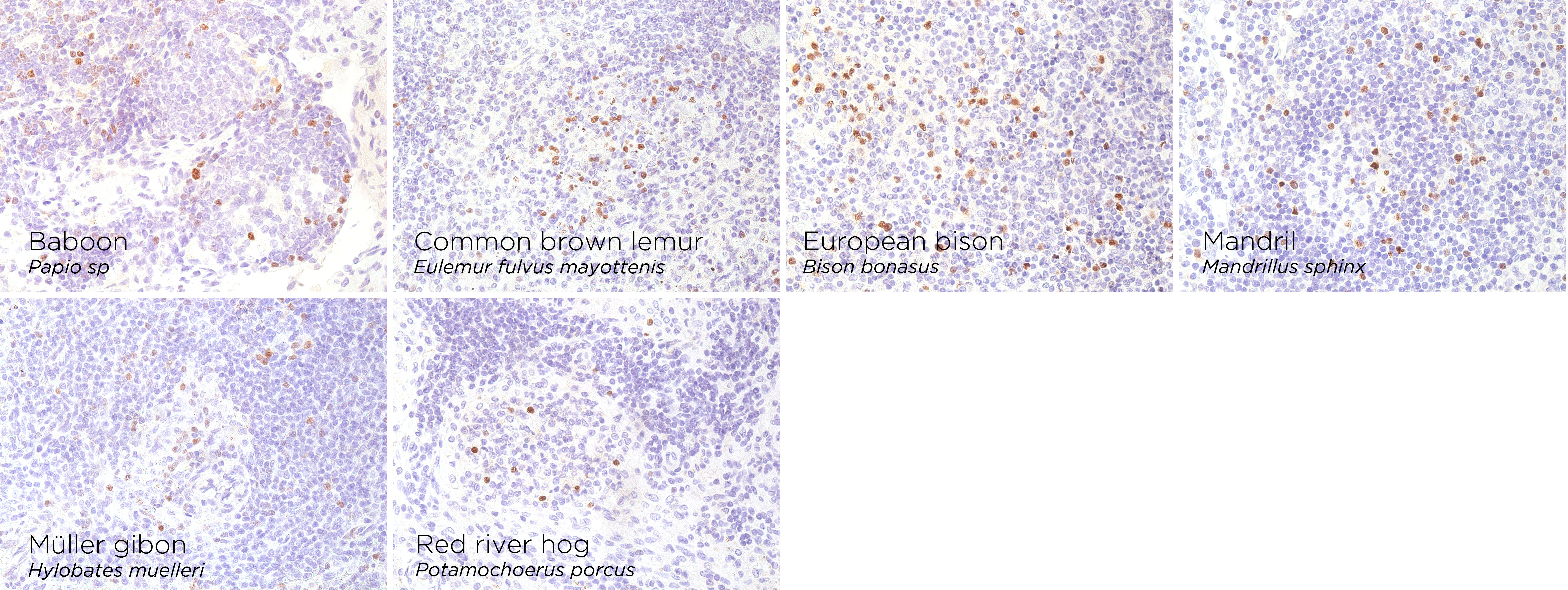
IHC-P Wild species
- African sacred ibis Threskiornis aethiopicus
- NV
- Asian black bear Ursus thibetanus
- -
- Baboon Papio sp.
- +++
- Badger Meles meles
- -
- Bush Dog Speothos venaticus
- Common brown lemur Eulemur fulvus mayottensis
- ++
- Emu Dromaius novaehollandiae
- NV
- European bison Bison bonasus
- +++
- Mandrill Mandrillus sphinx
- +++
- Otter Pteronura brasiliensis
- NV
- Patagonian mara Dolichotis patagonum
- NV
- Penguin Spheniscus demersus
- NV
- Raccoon Procyon lotor
- -
- Red flamimgo Phoenicopterus ruber
- NV
- Reindeer Rangifer tarandus
- -
- Rhea Rhea americana
- Sandbar shark Carcharhinus plumbeus
- Sitatunga Tragelaphus spekii
- -
- South American sea lion Otaria byronia
- Stork Ciconia ciconia
- NV
- Wild pig Sus scrofa
- NV
- Wolf Canis lupus