Antibody Identity card
Antibody information [ PDL-1 mouse specific]
GOYA536A
- Clone name GOYA536A
- Description Rat monoclonal
- Antigen used RBL1 cell line transfected with full lenght mouse PDL1 vector
- Epitope Unknown
- Isotype IgG2a
- Confirmed species reactivity Mouse
- Ab used Rat mAb clone name GOYA536A CNIO (tissue culture
| APPLICATION | Recommended concentration | Status | Protocol |
|---|---|---|---|
| Western blotting (WB) | 38μg/ml | Working | Western Blotting (WB) |
| Immunocytochemistry (ICC) | neat (tissue culture supernatant) | Working | Cytospin preparation technique |
| Immunohistochemistry (IHC-P) | Not tested | ||
| Immunoflourescence (IF) | neat (tissue culture supernatant) | Not tested | Immunofluorescence staining |
| Flow cytometry (FC) | 38 μg/ml | Working | Flow cytometry |
| IHC-P Species | Not tested |
Ab ID Card application validation / characterisation
WB Validation
Over-expression/cross reactivity

Gene inactivation
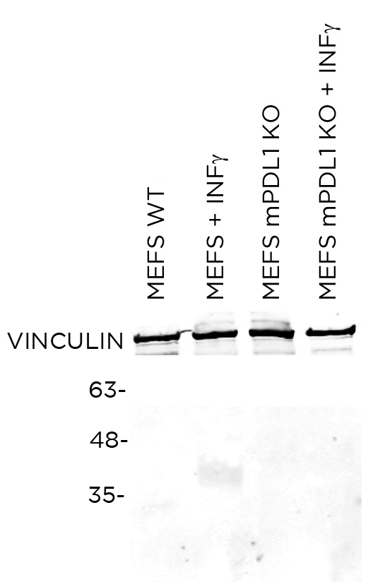
WB Characterisation
Endogenous expression

ICC / ICC-P Validation
Over-expression/cross reactivity
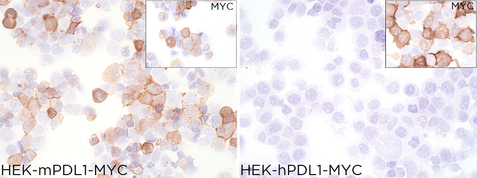
Gene inactivation
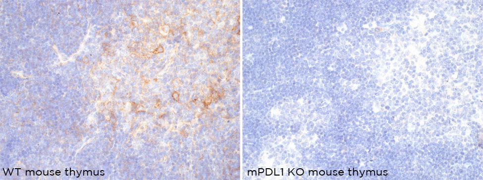
IHC-P Characterisation
Endogenous expression
Not tested
IF Characterisation
Endogenous expression

FC Validation
Over-expression/cross reactivity

Gene inactivation

FC Characterisation
Endogenous expression

IHC-P Domestic species
Not tested
IHC-P Wild species
Not tested